Information for Oncology Treatments
Here is some information on common questions we receive. Please note no two cases are exactly the same and information is general in nature.
Lymphoma
Lymphoma is the most common haematopoietic malignancy we see affecting dogs and cats. It is important to note that “lymphoma”, “lymphosarcoma” and “malignant lymphoma” are all the same condition and do not imply any difference in behaviour. It has been shown in humans with non-Hodgkin’s lymphoma that the lymphocyte becomes cancerous due to an accidental insertion of a proto-oncogene within the antibody-reading template during the response to an antigen. Research from genomic profiling has shown that the majority of dogs have deregulation in the mechanism that controls which genes are expressed. In short, this leads to silencing of anti-cancer genes, and an increase in the chance for mutations to occur. Further investigation into the genes involved is currently underway.
The success rate treating lymphoma is generally high. Around 85% of dogs enter remission on most protocols. Non-multicentric forms in dogs can still respond, but as a group do not perform as well with respect to remission rates. In cats, any form of lymphoma can respond well; around 2 in 3 cats enter remission whilst 1 in 3 are long-term survivors and often cured. Delaying treatment ofhigh-grade lymphoma, with or without prednisolone, worsens the prognosis.
Therapies range from 3 to 6 months of treatment, depending on the protocol. Costs can be significant, with each treatment visit being hundreds of dollars. Visits range from weekly (initially) for the intensive therapies, to monthly for the less aggressive protocols. The intention is that quality of life is normal during treatment as well as after, and this is realised for almost all patients.
Patients are readily referred to Perth Veterinary Oncology by on-line form, email, or phoning 9204 0400. Dr Ken Wyatt and Dr Jessica Finlay are the only Veterinary Oncologists in Western Australia.
Mast Cell Tumours
The introduction of the Kiupel grading scheme has improved the prediction of dogs at risk of death. All dogs classed as having a high Kiupel grade mast cell tumour should be considered candidates for adjuvant therapy.
In situations where appropriate margins (3cm in all directions) are not achievable or allowed, or even if surgery is not allowed at all, medical therapy is successful in many cases. Around one in five dogs with highly aggressive metastatic mast cell tumour (with no surgery performed) remain disease free after 2 years, compared to median survivals of a few weeks without any therapy.
Electrochemotherapy has been proven as successful as surgery in recent trials for tumours of less than 3cm diameter.
Finally, imatinib (Glivec), masitinib (Masivet) and toceranib (Palladia) have recently produced very encouraging results against metastatic and non-resectable disease. Results appear to be temporary when used as monotherapy but allow patients without good surgical options to receive chemotherapy with greater likelihood of success. When combined with cytotoxics therapy the potential for more durable results appears to improves markedly.
Approach:
Low Kiupel tumours with clear staging and good margins: monitoring only
Low Kiupel tumours with clear staging and incomplete margins: refer to Perth Veterinary Oncology for advice on electrochemotherapy
High Kiupel tumours or advanced disease: refer to Perth Veterinary Oncology for advice on medical therapy
Patients are readily referred to Perth Veterinary Oncology by on-line form, email, or phoning 9204 0400. Dr Ken Wyatt and Dr Jessica Finlay are the only Veterinary Oncologists in Western Australia.
Melanoma
Melanomas of the skin are generally benign in behaviour in both dogs and cats. In dogs, 15% of skin melanomas and 33% of nail bed melanomas behave in a malignant manner. Poodles also appear to be predisposed to the aggressive forms (~85% of poodle melanomas behave aggressively). Melanomas of the skin are best removed surgically. Rapid growth, high mitotic rate (greater than 3 per 10 high power fields), or an ulcerated surface predicts aggressive behaviour and tumours showing these characteristics should be removed with larger margins and referred. Intralesional/electro-chemotherapy can be effective in locations for which surgery is likely to cause problems, such as the oral cavity or distal limbs.
The majority of oral (non-lip) melanomas behave in a malignant metastatic manner. Grade is NOT predictive for outcome however those with low mitotic counts of less than 4 per 10 high power fields have a more favourable outcome.. Therapies include surgery, radiation therapy or intralesional/electro-chemotherapy. Surgery often needs to be aggressive with margins (i.e. hemimandibulectomy), however even with aggressive resection, the local recurrence rate is around 25%. Whilst median survival with surgery alone is around 8 months, at least a third of the dogs enjoy survival beyond 2 years. If surgery is not feasible or desired by the owner,radiation therapy or intralesional/electrochemo-therapy is successful in inducing remission in approximately 50% of patients. Median survival in responders is greater than 1 year.
Uveal melanomas in cats are generally malignant and require enucleation, whilst limbal melanomas are locally infiltrative and may be removed from the eye if diagnosed early.
Systemic chemotherapy is available for dogs with oral melanoma to control metastatic spread. Systemic chemotherapy is best reserved for melanomas with histopathologic or imaging evidence of aggressive behaviour. For those with reasonably good quality of life, therapy can be employed to maintain quality of life. Intralesional/electrochemo-therapy can be employed if the primary mass is limiting quality of life and surgery is not possible or declined by the pet’s owner.
Vaccine immuno-therapy is available for canine melanoma. It is administered by a needle-free device on 4 occasions at 2 week intervals, but otherwise is as readily and rapidly given as other vaccinations for dogs.
Patients are readily referred to Oncology at Perth Veterinary Specialists by on-line form, email, or phoning (08) 9204 0400. Dr Ken Wyatt and Dr Jessica Finlay are the only Veterinary Oncologists in Western Australia.
Haemangiosarcomas
Haemangiosarcomas occur most often in German Shepherd dogs (and large breeds in general). Most tumours in both dogs and cats occur in the spleen. However, it is important to note that in the dog, only around one half of splenic masses are haemangiosarcoma, and most of the remainder are benign. In many dogs with large spleens therefore, splenectomy is curative. However, canine splenic haemangiosarcoma has usually metastasised before recognition. The right atrium, liver, lungs, and brain are common sites. With splenectomy only, median survival times are typically ~3 months. Large samples from various regions of the tumour should be submitted for histopathology, as much of the gross appearance of splenic haemangiosarcoma may be haematoma surrounding the tumour. In addition, unrelated benign liver masses are common in aged dogs, and may appear similar to metastatic spread. It is therefore strongly advised to remove the spleen and biopsy lesions consistent with metastatic spread elsewhere, rather than euthanase based on gross appearance.
Cutaneous haemangiosarcomas occur most frequently in dogs with poor skin pigmentation. For dermal tumours, if the histopathology shows no invasion beyond the dermis with adequate margins, no further treatment is required.
Therapy depends on the stage of disease. Following surgery of the non-cutaneous forms, maximum tolerated doses of chemotherapy is recommended. Some dogs are now living well into their second year, with occasional long-term survivors. Staging should be preformed to obtain a base-line. As an alternative to MTD chemoterapy treatment, anti-angiogenesis treatment such as metronomic chemotherapy can be employed. Anti-angiogenesis treatment does not tend to produce remission but prevents growth of the cancer in some patients. Patients suitable for this form of therapy must therefore have good quality of life when commencing treatment (as there is no point in prolonging the life of an ill patient). Ideal therapy employs both systemic MTD chemotherapy concurrent with metronomic therapy.
What to do:
For cutaneous haemangiosarcoma, excisional biopsy is often enough. Refer if the lesion is large or there are too many lesions to remove.
For non-cutaneous haemangiosarcoma, either refer once histopathology results are back, or for the initial surgery if preferred. Full staging (imaging) is readily done at Perth Veterinary Specialists. A biochemical panel is of no direct necessity, other than as a usual pre-surgical precaution. A CBC will be needed – anaemia is common due to haemorrhage, erythrophagia or haemolysis (not immune mediated). Bleeding defects are possible and should be checked pre-operatively.
When to refer:
At any point from documentation of the lesion, to as soon as possible following surgery. Histopathologic confirmation of the tumour is required before chemotherapy can commence. Dogs with metastatic disease can still be treated, however with more short term risk.
Patients are readily referred to Perth Veterinary Oncology by on-line form, email, or phoning 9204 0400. Dr Ken Wyatt and Dr Jessica Finlay are the only Veterinary Oncologists in Western Australia.
Osteosarcoma
Osteosarcoma has metastasised in over 95% of patients before diagnosis. Thoracic radiographs are positive at the time of diagnosis in only 10% of patients. The remainder have metastases too small to be visible. The ideal osteosarcoma patient for chemotherapy is one with the primary tumour removed and no visible metastases. Therapy is commenced once histopathology results confirm the disease (if no biopsy pre-amputation) or once recovery from surgery is adequate to leave the patient systemically well (within 5 days). Treatment with surgery alone produces median survival times of around 3-4 months. Medical therapy allows most dogs another 10-12 months with around 20% going much longer. Osteosarcomas in regions other than the limbs are often still readily treatable with similar or better prognoses. For patients with early lesions in the distal radius, a limb-sparing procedure may be performed. This is a major surgical procedure with small but real problems of infection and local tumour recurrence, but is indicated when owners wish to preserve the limb at all costs. Radiation therapy is now also available.
Medical therapy alone is an option for patients unsuitable for amputation or radiation therapy. Chemotherapy is combined with zolendronate (bisphosphonate). Quality of life improves for most dogs for 6 to 12 months.
Recent data shows that some dogs with advanced metastatic disease can still have prolonged survival following standard therapy. Median survival is around 2 months so clearly most dogs still succumb rapidly. Therefore, euthanasia based on the presence of advanced disease will rob a small percentage of patients from extended survival. It is still humane to treat a dog with metastatic osteosarcoma in many cases given that amputation is often the most effective form of pain control, however short survival is expected.
What to do:
Biopsy or aspirate is wise pre-operatively; however if the bone lesion is so severe that amputation will be required regardless of the diagnosis, and the owner consents, amputation with histopathology is acceptable. The differential diagnostic list includes other tumours (usually haemangiosarcoma or fibrosarcoma), fungal infection, bacterial infection, and bone cysts. Chest radiographs (both laterals and a DV or VD view) or thoracic CT are worth performing and can be done at Perth Veterinary Specialists. A biochemical panel is of no direct necessity, other than as a usual pre-surgical precaution. However, if done, a normal ALP activity is a good prognostic sign.
When to refer:
At any point from radiographs showing a lytic & proliferative lesion in bone, to as soon as possible following amputation. Histopathologic confirmation of the tumour is ideal before commencing chemotherapy. Dogs with metastatic disease can still be treated, however with less emphasis on long term control. Metastasis to other bone sites carries a better prognosis than to the lungs.
Patients are readily referred to Perth Veterinary Oncology by on-line form, email, or phoning (08) 9204 0400. Dr Ken Wyatt and Dr Jessica Finlay are the only Veterinary Oncologists in Western Australia.
Carcinomas
Carcinomas are cancers derived from epithelial cells. Common examples such as mammary carcinomas and squamous cell carcinomas have been covered on other pages. This page details the remaining carcinomas, such as anal sac carcinomas, bronchial carcinomas, and GI tract carcinomas. These tumours will often metastasise, typically to local lymph nodes and lungs, amongst other sites. They have a moderate response to chemotherapy. Hence, surgery is the first treatment. In addition:
- Adjuvant chemotherapy reduces the microscopic burden remaining after surgery.
- Neoadjuvant therapy reduces the size of the tumour to allow surgical removal.
- Palliative chemotherapy improves quality of life and delays its progression.
- Intracavitary infusion of chemotherapy can have excellent success in controlling effusion.
- Intralesional injections of chemotherapy or electrochemotherapy can be very effective when surgery is declined; they have the advantage of avoiding systemic side effects, yet increasing local response rates versus systemic chemotherapy.
- Metronomic (frequent oral) chemotherapy and tyrosine kinase inhibitors can be employed to inhibit blood vessel development, slow growth and thereby maintain quality of life for an extended period.
Summary:
- Stage the disease. Ideally a combination of cytology of, regional nodes and imaging. CT is preferable over radiographs.
- Remove visible tumour (if possible) followed by referral to Oncology at Perth Veterinary Specialists for advice regarding adjuvant therapy.
- If curative intent surgery is not possible, refer to Oncology for advice on neoadjuvant or palliative treatment, or refer to Surgery.
- Costs vary greatly but are typically hundreds of dollars per chemotherapy treatment for most dogs and cats. Generally, from 1 to 6 treatments are given. Metronomic therapy costs a few dollars per day for most dogs.
Patients are readily referred to Perth Veterinary Oncology by on-line form, email, or phoning 9204 0400. Dr Ken Wyatt and Dr Jessica Finlay are the only Veterinary Oncologists in Western Australia.
SCC
Cats
Nasal, eyelids & ears:
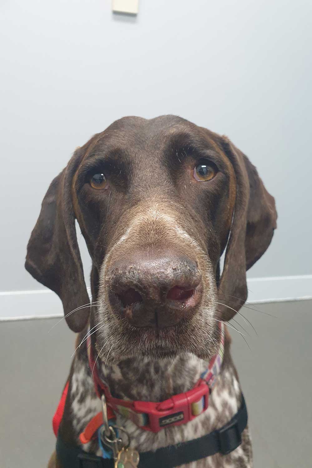
Pre-cancerous lesions appear as erythemic, scaley regions. Once a non-healing ulcer is present, the lesion should be presumed cancerous. Carcinoma <2mm diameter may be non-invasive until random mutations allow angiogenesis. It therefore should be treated as soon as possible to prevent progression. Simple cryotherapy and strontium radiation (not available) are both suitable for lesions of this size. Once larger, inappropriate treatment will delay definitive therapy and worsen the prognosis. This is the main reason for failure in the treatment of this condition. Larger lesion treatments include surgical resection of the entire rhinarium, spray application of liquid nitrogen onto the lesion and electrochemotherapy. Referral is encouraged for recurrent lesions, or ulcers 3mm in diameter or larger.
Oral
A SCC in this location may appear as a small proliferative mass or ulcer but is always aggressive. Radical surgery with tube feeding can provide most cats with a second year but is only suitable for committed owners. If radical surgery is declined, intralesional/electro-chemotherapy or radiation therapy can be performed. Most patients respond to treatment but do not enter remission; quality of life can be maintained for significant periods, however. Medical treatments are supportive and may be very helpful with palliative expectations.
Dogs
Ventral abdomen
The entire non-pigmented ventral body is typically affected. Surgery is usually the first choice, but must be aggressive with margins of at least 1cm. Referral is wise to plan measures to slow or prevent other lesions from developing. When surgery is not appropriate, referral for deep cryotherapy, or intralesional/electro-chemotherapy is advised.
Oral
A pilot study has shown excellent success with chemotherapy and piroxicam, with many dogs not even requiring surgery. Durable responses occurred even in the face of regional node involvement. The tonsillar form of the disease carries a poorer prognosis. Compounds such as toceranib have anecdotal support.
Patients are readily referred to Perth Veterinary Oncology by on-line form, email, or phoning 9204 0400. Dr Ken Wyatt and Dr Jessica Finlay are the only Veterinary Oncologists in Western Australia.
Soft Tissue Sarcomas
Soft Tissue Sarcomas are a group of neoplasms including:
Fibrosarcoma
Leiomyosarcoma
Haemangiopericytoma
Liposarcoma
Rhabdomyosarcoma
They generally behave in a similar fashion. The important considerations are:
- A pseudocapsule of compressed cancer cells often surrounds them, which may make them falsely appear to be readily ‘shelled out’ during surgery. Wide margins are required, which may require a radical procedure such as amputation if done with curative intent.
- The metastatic rate is generally around 25% or less, and typically to distant sites such as the lungs. Tumours classed as high grade, or undifferentiated/anaplastic, have much higher rates of metastasis.
- Measurable (visible) disease is often best treated by surgery.
The diagnostic investigation should start with a biopsy. Note that an unsuccessful attempt at excision (i.e. removing without first determining that wide margins are required) will reduce the potential for definitive surgery to succeed. Thoracic radiographs (2 laterals, 1 DV view) are wise prior to definitive excision with very wide and deep margins. Referral for adjuvant chemotherapy is indicated for high grade or anaplastic tumours. If definitive surgery is declined or not possible, other options include:
- Metronomic chemotherapy following marginal excision (without resection of overlying skin if necessary). Treatment inhibits angiogenesis and thereby delays recurrence. In a recent trial, dogs treated in this manner had tumour recurrence on average after 3 years (rather than 6 months with surgery alone). Metronomic chemotherapy is daily and for life, at a cost of a few dollars per day.
- Electrochemotherapy. Generally one to two treatments are required. Results are variable but can lead to reduction in tumour size and/or prolonged periods (1 year) of growth inhibition. No side effects other than local inflammation occur.
- Radiation therapy
Patients are readily referred to Perth Veterinary Oncology by on-line form, email, or phoning 9204 0400. Dr Ken Wyatt and Dr Jessica Finlay are the only Veterinary Oncologists in Western Australia.
Mammary Tumours – Cats
Almost all mammary tumours in cats are malignant and metastatic. Hormone receptors are much less frequent; however there does appear to be a relationship with the administration of progestogens. Surgery is more aggressive than in dogs, as the disease is more locally invasive, such that unilateral mastectomy is advisable. Chemotherapy is advisable in cats with tumours larger than 3 cm diameter (median survival without treatment is 6 months) and/or with confirmed nodal spread. If visible tumour remains anywhere, responses are poorer.
Patients are readily referred to Perth Veterinary Oncology by on-line form, email, or phoning 9204 0400. Dr Ken Wyatt and Dr Jessica Finlay are the only Veterinary Oncologists in Western Australia.
Mammary Tumours – Dogs
The literature states that risk is related to the hormonal status of the bitch, and determined by the first few heats. In reality it is uncertain – the strength of the studies to date have been variable, and therefore it is wise to consider that spaying MIGHT reduce the risk of mammary carcinoma in dogs. European studies have also shown that dogs that are lean at 12 months of age are at lower risk. Many tumours contain hormone receptors on their surface, suggesting that sterilising dogs with these tumours may slow their growth. The more aggressive the tumour is, the less likely it will have receptors. We therefore do not recommend desexing dogs with high grade mammary carcinomas if the intent is to slow the growth of the tumour. There may of course be other justifications relating to individual patients for desexing at the time of tumour removal.
In addition to removal of the tumour, regional lymph nodes should be aspirated, or removed for histopathology and three view thoracic radiographs taken. Immunohistochemistry may help to grade the tumour more accurately. Chemotherapy should be considered in those dogs with histopathologic confirmation of vascular invasion, tumours over 3cm in diameter, vimentin expression, OR nodal involvement. Without treatment, median survival time for these dogs is little more than 3-4 months. Alternatively, treatment using metronomic chemotherapy (at home, oral) to prevent blood vessel development in dogs with visible metastasis has the potential to delay tumour growth. Interestingly, 1 research group found marked benefit from the administration of DDAVP at the time of tumour removal, potentially through vascular disruption of micrometastatic foci.
Patients are readily referred to Perth Veterinary Oncology by on-line form, email, or phoning 9204 0400. Dr Ken Wyatt and Dr Jessica Finlay are the only Veterinary Oncologists in Western Australia.
Canine Prostatic Carcinoma
Almost all prostatic tumours in dogs are malignant, and most are carcinomas. Prostatic carcinomas in dogs are not influenced by androgens; in fact prostatic disease in an entire male is most likely non-neoplastic. Because castration prevents inflammatory prostatic disease, but not carcinoma, prostatic neoplasia is a likely explanation for prostatic enlargement in castrated males. The majority of these tumours will have spread by the time of diagnosis, most commonly via local extension to the bladder, iliac lymph nodes lungs and pelvic/lumbar vertebrae. The most common clinical signs are those associated with the urinary tract (haematuria, pollakiuria, stranguria).
Diagnosis can be achieved by cytology or histopathology or BRAF mutation detection. Ultrasound examination is very helpful in documenting the disease and can be used to guide aspirates or biopsies. Surgery may result in urinary incontinence and is not expected to be curative. It is most useful for dogs that have urgent problems such as urinary obstruction for which correction will immediately improve quality of life. Chemotherapy can be successful in relieving clinical signs, reducing the size of the tumour and providing a greater period of time for the dog. Cytotoxic treatment is combined with piroxicam. Without treatment most dogs are euthanased within weeks or a few months of diagnosis.
Patients are readily referred to Perth Veterinary Oncology by on-line form, email, or phoning 9204 0400. Dr Ken Wyatt and Dr Jessica Finlay are the only Veterinary Oncologists in Western Australia.


